I noticed that there were 3 searches on “what is neurological sequelae” today that led to this blog site. I’m thinking you didn’t find the answer, so I thought I might go into this a bit. “Neurological sequelae”, whoa, big phrase, huh? Let’s break it down. The secret to answering the question is to look up the word ‘sequelae’. Wikipedia covers it pretty well (I know, the instructors say not to use Wikipedia, but this is not to use as a reference in a paper, so….): http://en.wikipedia.org/wiki /Sequela. It states, “A sequela, (pronounced /sɨˈkwiːlə/, plural sequelæ) is a pathological condition resulting from a disease, injury, or other trauma”. Now you can understand that ‘sequelae’ could be neurologic, cardiologic, metobolic, etc, right?
Of course to answer the question, you must know what the original disease, injury or other trauma is. For example, herniation would be one neurological sequela to increased ICP. Increased ICP could be a neurological sequela to a subdural hematoma. In other words, of a certain neurological problem, what could it progress into.
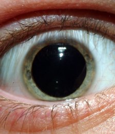
In Case You Don’t Know: Subdural hematoma – a hematoma (internal bleeding) between the dura (the inner most ‘covering’ layer around the brain) and the brain. ICP or Intracranial Pressure – pressure within the cranium or head caused by swelling of the brain or bleeding in the cranial cavity (this is not a good thing, because, once your fontanelle’s grow together as a baby, your head doesn’t really expand; it is a closed cavity). Herniation of the brain – if you have swelling of the brain or bleeding that is taking up space in the head, the brain, being crowded, only has one place to go; down the brain stem. This is known as the brain herniating (This can be partial or full. Full is rarely, if ever, recoverable and is assessed as a blown pupil):
Blown pupil:
If you have a question about specific neurological sequela, let me know what it is. I’ll see what I can figure out for you.
I love neurology, so give me all the questions you have.

Hi there, Im am currently working in Neurology in New Zealand as a student nurse. I am quite stuck at the moment and was wondering if you could help me out? I have to do case study and I am doing a 75 year old man who has an R) frontal acute on chronic sundural hematoma however no known falls, but he has a stoke many years ago and is on aspirin…what is the links between this? And I cant seem to find any info on what the definistion of an acfute on chronic subdural is.. new bleed on an old bleed?
thanks heaps
k
Karina,
Look up stroke: https://health.google.com/health/ref/Stroke. Now, strokes cause damage, right? Possibly the blood vessel that broke causing the subdural hematoma (SDH) was damaged during the stoke but not damaged enough to cause a bleed at the time of the stroke. We know aspirin thins the blood making any bleed worse by prolonging the clotting time.
Now, let’s look at the classifications of SDH; acute, subacute, and chronic. Read this from emedicine: “Generally, acute SDHs are less than 72 hours old and are hyperdense compared with the brain on CT scan. Subacute SDHs are 3-20 days old and are isodense or hypodense compared with the brain. Chronic SDHs are 21 days (3 wk) or older and are hypodense compared with the brain. However, SDHs may be mixed in nature, such as when acute bleeding has occurred into a chronic SDH.” – http://emedicine.medscape.com/article/247472-overview. So, we can deduce from this that the man had a chronic SDH and now in addition to that, he has a new additional bleed. Actually, the emedicine article I just cited is a great reference for your whole case. Take a look at the part about brain atrophy in the elderly.
Good luck with your case and all your classes!
Amy
MRI report of One Of Our Relatives shows Impressions as Folows:
Symmetrical Areas Of Altered Signal Are Seen In The Caudate And Lentiform Nuclei, Corona Radiata And Centrum Semiovale Of Both Cerebral Hemispheres As Well As In The Cerebellar White Matter In A Pattern Suggestive Of Sequelae Of Global Hypoxia. Small Foci of Old Petechial Hemorrhage Are Seen In The Left Frontal And Occipital Cortex And The Right Thalamus Repectively…
Please let me know in simple terms What Does It Mean?
Also, what are the chances of Recovery?
Is There Any Treatment Available For This?
I’m sorry, Ravinder, that this has happened to a relative. But this is really something you need to be discussing with your relative’s Doctor, especially as the MRI is not the whole picture. Thoughts are with you and your family.
Thanks amy47… But i would be highly obliged to you if you could please throw a light on this report…
Doctor’s are always positive… Until the last breath, they always keep you on assuring that everything will be fine…
Same is the case with my relative’s doctor…
He says, patient will heal with time.. But he is not sure about the time its gonna take…